Endoscopic Rhizotomy
Endoscopic Rhizotomy
Endoscopic rhizotomy is a minimally invasive endoscopic surgery that allows direct visualization of the medial branch nerve that supplies the facet joints in the back of the spine. The surgery takes the percutaneous RF facet denervation procedure an important step further by providing direct endoscopic visualization of the posterior spinal anatomy and nerves. An incision that is less than a quarter of an inch is made, and a camera is inserted in the spine. By cutting a section of the medial branch nerve, the pain signal is interrupted. This surgery can be performed on the cervical, thoracic, and lumbar spine. It can also be performed on the sacroiliac joint for sacroiliac joint disease.
Medial branch nerves are very small nerves that innervate the facet joints of the spine. Facet joints are the joints connecting the different vertebra of the spine to each other. The joints are present on both sides of the spine from the neck to the lower back.
When is Endoscopic Rhizotomy indicated?
Radiofrequency Rhizotomy is indicated if a diagnostic procedure, called a medial branch block, is successful in confirming the patient’s back pain is originating from the facet joints. For the sacroiliac joint, a successful sacroiliac joint injection is needed.
Medial branch block is a procedure where local anesthetic is directly placed near the medial branch nerve to block the pain signal carried from the facet joints to the brain. It is a diagnostic tool and typically provides only temporary relief from pain. It is critical in assisting spine specialists in diagnosing the specific cause of your back pain.
After the medial branch block your pain may:
- Go away for a few hours
- Go away for a few days
- Not reduce at all
If the pain is relieved after the medial branch block, this indicates that the origin of the pain are the medial branch nerves that were numbed. At that point, we would likely recommend a radiofrequency ablation or an endoscopic rhizotomy to relieve the pain for a longer period of time. With the radiofrequency technique, the nerves regenerate over time and the pain returns after a few months. With the endoscopic technique, a section is cut from the nerve, preventing the nerve from being able to regenerate.
How is an Endoscopic Rhizotomy performed?
Endoscopic rhizotomy is an outpatient, same day, true minimally invasive surgery. During the day of your procedure, you will be taken to the pre-op area where trained nursing staff will get you ready by taking vitals and reviewing your medications. Your blood sugar and coagulation status may also be checked if needed. Then you will enter the operating room where you will lie face down on a table for treatment of the painful area.
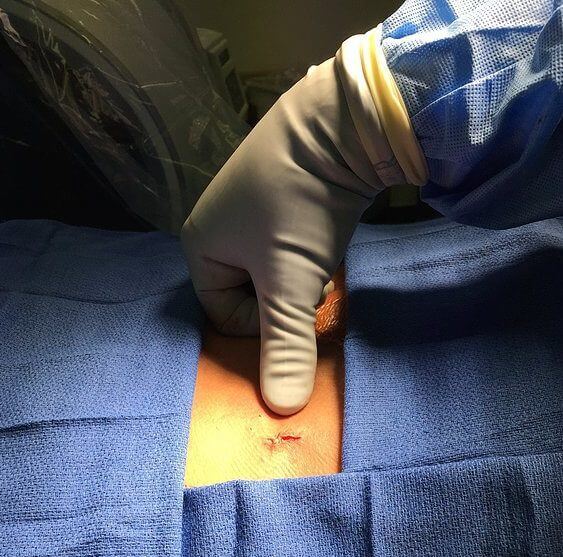
The surgery is performed under deep sedation so there is no pain during the surgery. A small (7mm) incision is made in the surgical area and an endoscopic cannula with a camera is inserted into the spine. The doctor is guided by fluoroscopic X-ray to place the camera in the correct position.
The camera allows the surgeon to see the inside of your spine where the nerve usually resides. The surgeon utilizes a microscopic cauterizing instrument to find the small nerve branches that supply the joints in the spine. After identifying the nerve, a section is cut from the nerve, preventing any regrowth in the future.
The camera is removed and the incision is closed with a single absorbable suture that is buried under the skin, so that no suture removal is needed. The procedure takes about 45 minutes to complete but may take longer depending on the number of nerves required to be treated.
What are some possible risk or complications from this minimally invasive surgery?
With the use of careful imaging which allows direct visualization of the spine and spine specialsits trained in the latest endoscopic techniques, complications are very rare. But with all medical procedures, complications may occur. To help minimize risks please follow all directions given to you by your physician. Ensure that all your treatment options are explained so you are aware of the risks and benefits of this surgery.
Some complications may include:
- Infection: Your surgeon cleans and sterilizes your back before every surgery to prevent this from occurring. This risk is minimized as you are given IV antibiotics one hour prior to the start of surgery.
- Increased pain may occur after the procedure in the area where the camera was placed. This may last up to a week or rarely longer. However, it typically resolves completely.
- You may also experience an area of numbness, usually in the area which was originally painful.
- The procedure may take up to six weeks for complete benefit, so improvement may not be immediate.
You will need someone to drive you home once you are discharged. As always, follow the instructions of your care provider and have your questions answered prior to the procedure.
One comment
Pingback: Everything You Need to Know Before Going for a Rhizotomy – Site Title