Monday - Friday (BY APPT ONLY)Saturday and Sunday - CLOSED718-962-7246admin@anagenesisspine.com78 Todt Hill Rd, Suite 112Staten Island, NY 10314 New York, NY

Latest news

Classic list

Globally incubate standards compliant channels before scalable benefits. Quickly disseminate superior deliverables whereas web-enabled applications.
pain-management.jpg

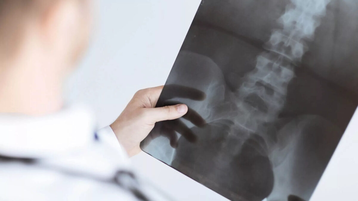
Pain Management Doctors in Staten Island

A king’s ransom to whoever has been pain-free their entire life. Even though most of us dread pain in any form, this unpleasant sensation is in fact a normal and, in many cases, a desired warning mechanism.


What is pain and why is it good (sometimes)?

According to the International Association for the Study of Pain, pain is: “an unpleasant sensory and emotional experience associated with actual or potential tissue damage”. In other words, pain, although an uncomfortable feeling, serves as a warning mechanism that tells the body something might be wrong.

In order for the warning mechanism to work, our organisms must be equipped with highly sensitive nerves that detect potentially dangerous changes in pressure, temperature, or chemical balance and send information about these to the brain.

Imagine a situation where you are about to touch an extremely hot pot lid. As you extend your hand and begin to grip the lid, the nerves on your fingertips (peripheral nervous system or PNS) immediately alert your brain (central nervous system or CNS) that the temperature of the object is so high that it can cause damage to the tissue. The brain then commands your hand to halt the action right there, in order to prevent the injury. You involuntarily withdraw your hand and drop the lid. It all takes a fraction of a second and prevents the tissue – your skin – from getting burned.

Pain is a complex and sophisticated protective mechanism. That being said, we must remember that sometimes the body’s alert systems don’t work properly or that an injury led to damage so severe that the body is in a prolonged or constant state of pain.

Such a situation is abnormal and one should always consult a pain management doctor without delay. This can prevent further, potentially irreversible, damage to the body and ease the emotional distress that comes from living in excruciating pain.


Pain is personal

Pain is a very subjective feeling. That means that people experience pain, even the same type of pain, differently, and no one can know exactly what somebody else’s pain feels like. Because everyone can experience pain in a way unique to them, diagnosis and treatment of pain is a challenge to pain doctors.


Many pain clinics focus on treating symptoms and relieving pain. In contrast, at Anagenesis Spine & Pain Medicine, our pain doctors in Staten Island practice an individualized approach: they concentrate on identifying the source of pain and then determining the most effective and beneficial treatment plan for each particular patient.


What are the different types of pain?

There are several ways to categorize pain but, in most cases, it can be classified as one of two kinds:

  1. Acute pain: Acute pain occurs suddenly, for example coming immediately after damage to tissue such as bone, muscle or organs, but it has a limited duration. Once the underlying cause of pain is treated and the injury heals, the pain stops. However, if the injury doesn’t heal well and the pain persists, then it may become classified as chronic.
  2. Chronic pain: With chronic pain, your body continues to send pain signals to your brain, even after the injury heals. This can last from several weeks to years. Headache, postsurgical pain, post-trauma pain, lower back pain, cancer pain and arthritis pain are examples of chronic pain types. Chronic pain is also associated with conditions such as fibromyalgia or osteoarthritis and may be very difficult to manage using standard pain management modalities.

Pain can also be classified by the kind of damage that caused it:

  • Nociceptive pain is caused by the stimulation of nociceptors, which are pain receptors distributed throughout the body. When the nociceptors are stimulated by a cut, tear, burn or fracture, they send electrical signals to the brain (“alarm the brain”) causing us to feel pain. Most types of injury or inflammation result in nociceptive pain.
  • Neuropathic (nerve-based) pain results from damage to or dysfunction of the nervous system (central or peripheral) caused by accidents, injury, disease, or infection. Neuropathic pain is not the direct result of any event or injury, but the body sends unprompted pain signals to the brain, anyway.

Our pain management doctor in Staten Island knows that recognizing the right kind of pain is vital for successful pain management. Knowing what kind of pain the patient is experiencing helps to understand the idiosyncrasies and challenges of the particular case and plays an important part in providing the most effective, and adequate treatment.


What is Pain Management

The American Board of Pain Medicine defines pain medicine/pain management (also referred to as algiatry) as a branch of medicine that is concerned with the prevention of pain, and the evaluation, treatment, and rehabilitation of persons in pain.

Modern pain management doctors – including our Staten Island pain medicine practice – employ an interdisciplinary approach for easing the suffering and improving the quality of life of those living with all types of pain.


The purpose of Pain Management

Pain, and especially chronic pain, may negatively impact a person’s everyday function. Personal, social and professional life may be negatively affected by both acute and chronic pain, regardless of its cause. Stress and emotional trauma associated with experiencing debilitating pain may further impede recovery from the injury or disease.

Proper pain management results in faster recovery and significantly enhances a person’s quality of life. On the other hand, poorly managed pain may become a syndrome on its own and cause a downturn in a person’s emotional and physical health.

Sometimes it is impossible to eliminate pain completely and in such instances, our pain management doctors in Staten Island turn their focus to minimizing pain. By making pain bearable, and improving functions of body parts affected by pain, patients can regain physical independence and get back to their daily routines, activities and professional life.


Pain Management Procedures

At Anagenesis Spine & Pain Medicine, our Board Certified Spine & Pain doctors in Staten Island offer the most comprehensive pain management programs combining different modalities to help alleviate pain. Depending on your specific condition, our doctor will discuss with you the following procedures:

  • Interventional Treatments
    • Cervical Epidural Steroid Injection
    • Lumbar Epidural Steroid Injections
    • Discography
    • Facet Joint Injections
    • Medial Branch Blocks
    • Radiofrequency Ablation
    • Sacroiliac Joint Injection
  • Regenerative Medicine
    • Platelet Rich Plasma Injection
    • Stem Cell Procedures
  • Same Day Surgical Treatment
    • Endoscopic Discectomy
    • Endoscopic Rhizotomy
    • Kyphoplasty


Learn more about Pain Management in Staten Island

There is no need to live in chronic pain!

We treat every patient as an individual who requires specialized care. We take time to listen to our patients, to get to know the conditions they suffer from inside and out, to know their everyday life challenges.

Only this approach allows us to properly diagnose and treat pain. Rest assured that each procedure and treatment plan will be explained and discussed with you in detail.

Anagenesis and Spine & Pain Medicine is your pain management clinic in Staten Island. Your well-being is our priority. Our pain specialists always focus on the correct diagnosis, and the most effective and, if possible, the least invasive procedures. Contact us today at 212 – 235 – 1265.


The material contained on this site is for informational purposes only and DOES NOT CONSTITUTE THE PROVIDING OF MEDICAL ADVICE, and is not intended to be a substitute for independent professional medical judgment, advice, diagnosis, or treatment. Always seek the advice of your physician or other qualified healthcare providers with any questions or concerns you may have regarding your health.


kyphoplasty-1200x1200.jpg

What is a Kyphoplasty?

Balloon Kyphoplasty is a minimally invasive procedure for the treatment of vertebral compression fractures, commonly called spinal fractures. This type of fracture is often caused by conditions that affect bone mass such as osteoporosis, cancer, or long-term use of oral corticosteroids. Spinal fractures cause the vertebral body to crack or collapse, altering the shape of the spinal column. Just one fracture can change the vertical alignment of the spine and can lead to additional fractures.

It has been estimated that over 700,000 spinal fractures occur annually in the U.S. alone. Significant pain and some degree of kyphosis is common, especially in patients suffering from multiple fractures.

Before minimally invasive surgery, the only treatment option for patients with spinal fractures was open surgery. Outcomes were poor, primarily because attaching hardware to bone that is already “soft” or compromised is difficult and not successful. Balloon Kyphoplasty is an innovative technique that can restore the vertebra to a near-normal shape, thereby maintaining spinal alignment. Before the procedure, the patient usually undergoes imaging studies to pinpoint the location of the fracture. Many patients report immediate relief from pain after the procedure due to the exothermic reaction of the bone cement hardening process.


What is a lumbar epidural steroid injection?

During this procedure, a corticosteroid (anti-inflammatory medicine) is injected into the epidural space to reduce inflammation and pain. Your physician may inject into the epidural space from behind, this is called an interlaminar injection. When your physician goes in from the side, it is called a transforaminal injection. If the needle is positioned next to an individual nerve root, it is called a selective nerve root block. When performed from below it is called a caudal injection.


What does a Kyphoplasty procedure entail?

Through two small incisions on either side of the spine, the spine specialist inserts a trocar into the fractured vertebra under fluoroscopic guidance. One to two small orthopedic balloons are passed through the trocar and inflated inside the collapsed vertebral body. Inflation of the balloons raises the collapsed vertebral body in an attempt to restore normal anatomy.

Once height and shape of the vertebra has been restored, the balloons are deflated and removed. The “hollow” or void created by the balloons are then filled with bone cement. The bone cement hardens quickly, creating an internal cast thus stabilizing the fracture.


Do I need to be in the hospital for a Kyphoplasty?

Balloon Kyphoplasty may require an overnight hospital stay, depending on medical necessity. This procedure is typically performed on an outpatient basis. The procedure takes about one hour per fracture level treated and many patients are able to resume normal activity rather quickly.


endoscopic-rhizotomy.jpg

Endoscopic Rhizotomy

Endoscopic rhizotomy is a minimally invasive endoscopic surgery that allows direct visualization of the medial branch nerve that supplies the facet joints in the back of the spine. The surgery takes the percutaneous RF facet denervation procedure an important step further by providing direct endoscopic visualization of the posterior spinal anatomy and nerves. An incision that is less than a quarter of an inch is made, and a camera is inserted in the spine. By cutting a section of the medial branch nerve, the pain signal is interrupted. This surgery can be performed on the cervical, thoracic, and lumbar spine. It can also be performed on the sacroiliac joint for sacroiliac joint disease.

Medial branch nerves are very small nerves that innervate the facet joints of the spine. Facet joints are the joints connecting the different vertebra of the spine to each other. The joints are present on both sides of the spine from the neck to the lower back.

When is Endoscopic Rhizotomy indicated?

Radiofrequency Rhizotomy is indicated if a diagnostic procedure, called a medial branch block, is successful in confirming the patient’s back pain is originating from the facet joints. For the sacroiliac joint, a successful sacroiliac joint injection is needed.

Medial branch block is a procedure where local anesthetic is directly placed near the medial branch nerve to block the pain signal carried from the facet joints to the brain. It is a diagnostic tool and typically provides only temporary relief from pain. It is critical in assisting spine specialists in diagnosing the specific cause of your back pain.

After the medial branch block your pain may:

  • Go away for a few hours
  • Go away for a few days
  • Not reduce at all

If the pain is relieved after the medial branch block, this indicates that the origin of the pain are the medial branch nerves that were numbed. At that point, we would likely recommend a radiofrequency ablation or an endoscopic rhizotomy to relieve the pain for a longer period of time. With the radiofrequency technique, the nerves regenerate over time and the pain returns after a few months. With the endoscopic technique, a section is cut from the nerve, preventing the nerve from being able to regenerate.


How is an Endoscopic Rhizotomy performed?

Endoscopic rhizotomy is an outpatient, same day, true minimally invasive surgery. During the day of your procedure, you will be taken to the pre-op area where trained nursing staff will get you ready by taking vitals and reviewing your medications. Your blood sugar and coagulation status may also be checked if needed. Then you will enter the operating room where you will lie face down on a table for treatment of the painful area.

The surgery is performed under deep sedation so there is no pain during the surgery. A small (7mm) incision is made in the surgical area and an endoscopic cannula with a camera is inserted into the spine. The doctor is guided by fluoroscopic X-ray to place the camera in the correct position.

The camera allows the surgeon to see the inside of your spine where the nerve usually resides. The surgeon utilizes a microscopic cauterizing instrument to find the small nerve branches that supply the joints in the spine. After identifying the nerve, a section is cut from the nerve, preventing any regrowth in the future.

The camera is removed and the incision is closed with a single absorbable suture that is buried under the skin, so that no suture removal is needed. The procedure takes about 45 minutes to complete but may take longer depending on the number of nerves required to be treated.


What are some possible risk or complications from this minimally invasive surgery?

With the use of careful imaging which allows direct visualization of the spine and spine specialsits trained in the latest endoscopic techniques, complications are very rare. But with all medical procedures, complications may occur. To help minimize risks please follow all directions given to you by your physician. Ensure that all your treatment options are explained so you are aware of the risks and benefits of this surgery.

Some complications may include:

  • Infection: Your surgeon cleans and sterilizes your back before every surgery to prevent this from occurring. This risk is minimized as you are given IV antibiotics one hour prior to the start of surgery.
  • Increased pain may occur after the procedure in the area where the camera was placed. This may last up to a week or rarely longer. However, it typically resolves completely.
  • You may also experience an area of numbness, usually in the area which was originally painful.
  • The procedure may take up to six weeks for complete benefit, so improvement may not be immediate.
    You will need someone to drive you home once you are discharged. As always, follow the instructions of your care provider and have your questions answered prior to the procedure.

endoscopic-discectomy.jpg

What is an Endoscopic Discectomy?

Endoscopic discectomy is a minimally invasive spine surgery performed to treat disc problems that cause back and leg pain. A unique feature of this technique is the technology used to access the spine; through a keyhole (8mm) incision, surgery is performed using a specialized WOLF endoscope.

A WOLF endoscope is a specialized, german engineered, device with a tiny camera at the end. The endoscope delivers the surgeon an excellent view of the disc and related structures through this tiny incision. This enhanced view allows the surgeon to select the part of the disc to be removed that is causing your pain.

Instead of cutting through tissues (eg, muscles, ligaments) to access the spine, a dilator will gently separate soft tissue and are graduated in size to enlarge the operative field.

What are some of the benefits of Endoscopic Discectomy?

Some of the benefits of a true, minimally invasive, WOLF endoscopic discectomy include:

  • Tiny incision
  • Less tissue disruption
  • Less blood loss
  • Less post-operative pain
  • Accelerated recovery
  • Same Day Surgery
  • Can be performed under light sedation (minimizing anesthesia risk)


What conditions can Endoscopic Discectomy help with?

endoscopic discectomy

(DDD), disc herniation, and disc bulge are examples of disc-related problems that may compress or pinch spinal nerves and cause low back, buttock and leg pain. Other neurologic symptoms may include numbness, tingling, and weakness in one or both legs. The goal of endoscopic discectomy is to decompress the nerves, relieve symptoms, and enable the patient to quickly return to regular activities of daily living.

Not every patient with DDD or disc problem requires spine surgery. Dr. Kountis may recommend endoscopic discectomy for the following reasons:

  • Non-operative treatment fails to relieve symptoms
  • Progressive worsening of neurologic symptoms or pain, and/or neurological problems
  • Numbness, loss of function, and weakness (signs of neurologic dysfunction)


How is Endoscopic Discectomy performed?

Endoscopic discectomy is performed under local and/or intravenous anesthesia; the patient is awake during the surgery. The back area is cleansed and prepared for surgery. A small needle is inserted into the disc guided by fluoroscopy; a type of x-ray used during surgery. A tiny skin incision is made and the dilation tube is slipped into place followed by the endoscope.

Special miniature-sized instruments (eg, forceps, rongeurs) are advanced through the endoscope. A YAG laser may be utilized to remove only the damaged disc part and to bind the disc wall (annulus fibrosus). The laser also reduces (shrinks) the size of the disc to decompress nerve roots. The procedure takes about 30-minutes to one-hour per disc. The incision is closed with one suture and a small bandage.


platelet-rich-plasma-and-stem-cell-injection-1200x654.jpg

What are Stem Cells?

Regenerative medicine takes advantage of our natural ability to heal ourselves by using the healthy adult stem cells found throughout the body. Laboratory and clinical research has shown that it is possible to use adult stem cells to restore lost, damaged, or aging cells to effectively regenerate tissue and provide some patients with an alternative to surgery. Regenerative therapies are showing promise in orthopedic medicine, wound care, nerve restoration, and a variety of cardiovascular, neuromuscular, and autoimmune conditions.

Adult stem cells were discovered over 40 years ago when researchers found that cells derived from bone marrow had the ability to form various tissues. Adult stem cells are early stage cells that, under the right conditions, are capable of developing into other types of cells and hold the potential to regenerate damaged tissue.


Who is a Good Candidate for Stem Cell Injections?

The first step is to determine if you are a good candidate for an adult stem cell procedure. Your physician will want a history of your injury and a physical examination along with any x-rays, and an MRI. While stem cell therapy may be appropriate for certain conditions, it is not applicable for every condition. However, it is has proven to be a viable option for several individuals suffering from pain. Good candidates for adult stem cell treatment usually are:

  • A patient that wants to avoid the inherent complications that come with an invasive surgical procedure.
  • A pain sufferer that is interested in an option that offers potential for an overall improved quality of life.
  • A person who understands that surgery sets off a cascade of further degeneration, and is prepared to benefit from the body’s own regenerative mechanisms.

Every patient is different, the success of stem cell therapy is dependent on the severity of your condition and your body’s response to stem cell therapy.


What does the Procedure entail?

An adult stem cell procedure harnesses’ and amplifies the body’s natural mechanism for healing and anti-inflammation. Once you have been identified as a good candidate for the procedure, a member of our team will review the procedure with you and answer any questions that you may have. A brief overview of the procedure is below:

This mechanism uses adult autologous stem cells, derived from your own bone marrow.
In the procedure, the physician will aspirate these cells from your hip, concentrate them, then deliver them back into your body in the area of damage or injury to aid in natural healing.

In all, the process typically takes less than 45 minutes and the concentration of the cells takes about 12-14 minutes.
Because your procedure will utilize a concentrated serum of your own cells, the procedure is considered “autologous point-of-care.”

Once the procedure is complete, our staff will allow you to rest and will create a customized personal rehabilitation program for recovery. We will either ask you to come back for a few post-operative appointments or follow up with you by phone, email, or mail so we can track you healing progress.


What are the Potential Applications for Stem Cell Injections?

The spine is comprised of a team environment working together to provide function and mobility to the body. If an area of the neck or back is injured, the entire structure begins to compensate for the injury causing stability and function to decline. Creating the need to relieve this pain:

Possible conditions for treatment are: discogenic back pain, facet arthritis and degenerative disc disease.

Your shoulder is involved in small and large movements throughout the day. Shoulder injuries can be the result of the tiniest movements from writing to driving a car. Most people do not even know they are suffering from a shoulder injury. Once the injury is identified, the pain can become intolerable. Creating the need to relieve this pain:

Possible conditions for shoulder treatments are partial rotator cuff tears, labral tears, and mild to moderate osteoarthritis

The knee joint is used in almost all motion and over time will begin to degenerate from overuse. Knee pain can be extremely painful and challenging for anyone suffering from joint pain. Creating the need to relieve this pain:

Possible conditions for knee treatment are osteoarthritis, partial ligament tears: ACL; PCL, partial meniscal tears, and augmented ACL or PCL reconstruction

Foot and ankle injuries often occur. Over time, we collect scars making it difficult for our body to heal on its own. The constant pressures we cause with simples, line walking, or stretching, often exasperates the injury. Creating the need to relieve this pain:

Possible conditions for treatment: are mild to moderate osteoarthritis, tendon inflammation, partial achilles tendon tear, and muscle strain/sprain.

The constant motion of walking, running, biking or climbing involves our hips. The compression of these movements starts to wear on all our joints over time. In addition, the hips have a tendency to carry much of the burden on our body’s motions and pain. Creating the need to relieve this pain:

Possible conditions for treatments are osteoarthritis, labral tears, articular cartilage injuries and congenital deformities of the hip.


platelet-resized-1200x654.jpg

What is Platelet Rich Plasma?

PRP is produced from a person’s own blood. It is a concentration of one type of cell, known as platelets, which circulate through the blood and are critical for blood clotting. Platelets and the liquid plasma portion of the blood contain many factors that are essential for the cell recruitment, multiplication and specialization that are required for healing.

After a blood sample is obtained from a patient, the blood is put into a centrifuge, which is a tool that separates the blood into its many components. Platelet rich plasma can then be collected and treated before it is delivered to an injured area of bone or soft tissue, such as a tendon or ligament.

PRP is given to patients through an injection, and ultrasound guidance can assist in the precise placement of PRP. After the injection, a patient must avoid exercise for a short period of time before beginning a rehabilitation exercise program.


Is PRP Treatment Effective?

Several basic science studies in animal models suggest that PRP treatment can improve healing in soft tissue and bone. For example, increased numbers of cells and improved tendon strength have been noted in Achilles tendon injuries, and improved muscle regeneration has been shown in gastrocnemius (calf) muscle injuries.

These favorable findings in animal models have led to the widespread use of PRP treatment for a variety of conditions, including acute and chronic tendon problems, as well as injuries to ligaments and muscles. Some early-stage clinical studies in humans have been promising, but are limited by their study design and few patients.

The most promising early results have been seen when PRP treatment is used for chronic tendon conditions, such as lateral epicondylitis (tennis elbow) and Achilles tendinosis, which impacts the Achilles tendon.

In a small study involving knee osteoarthritis, PRP treatment was shown to be more effective than hyaluronic acid treatment. PRP has also resulted in positive or similar results when used in the treatment of rotator cuff tears and medial collateral ligament (MCL) injuries in the knee.

Overall, there is support of PRP treatment in published clinical studies. However, because PRP is created from a patient’s own blood, it is considered a relatively low-risk treatment with the potential to improve or speed healing.


What are some concerns regarding PRP?

Because PRP is given in the hopes of optimizing the initial inflammatory response of healing, anti-inflammatory medications should likely be stopped at the time of PRP treatment.

Also, PRP does contain endogenous growth factors, so some agencies consider it to be a performance-enhancing substance. For instance, the World Anti-Doping Agency and the United States Anti-Doping Agency forbid the injection of PRP within muscles because of the possibility that the growth factors could enhance a person’s performance. However, there are currently no data to suggest that PRP is actually a performance-enhancing substance. Major professional sports leagues have not yet addressed the topic of PRP.


Key Points to Remember

  • Platelet Rich Plasma (PRP) comes from a patient’s own blood.
  • PRP is a concentrated source of growth factors and cellular signaling factors that play a significant role in the biology of healing.
  • Basic science studies show that PRP treatment may improve healing in many tissues.
  • Anti-inflammatory medicines should be stopped before and after PRP treatment is given.

sacroiliac-joint-injection-1200x675.jpg

What is Sacroiliac Joint Pain?

Sacroiliac joint pain or SI joint dysfunction is the result of too much or too little motion occurring at the sacroiliac joint, which leads to inflammation and pain, which can often be debilitating. Part of the pelvis, the SI joint is attached to the front and back by the muscles and ligaments which surround it. Any of these can be the source of pain in a dysfunctional SI joint.


What are some symptoms of Sacroiliac Joint Pain?

Pain can range from dull aching to sharp and stabbing and increases with physical activity. Symptoms also worsen with prolonged or sustained positions (i.e., sitting, standing, lying). Bending forward, stair climbing, hill climbing, and rising from a seated position can also provoke pain. Pain is reported to increase during menstruation in women and sexual intercourse. Some examples include:

  • Lower back & buttock pain
  • Sciatic leg pain
  • Groin pain & hip pain
  • Knee Pain
  • Transient numbness
  • Urinary frequency (rare)


How is Sacroiliac Joint Pain Diagnosed?

Certain physical exam findings correlate with SI joint pain and dysfunction. The current “gold standard” for diagnosis of sacroiliac joint dysfunction emanating within the joint is a sacroiliac joint injection confirmed under fluoroscopy or CT-guidance using a local anesthetic solution.


How do you treat Sacroiliac Joint Pain?

Treatment is often dependent on the duration and severity of the pain and dysfunction. In the acute phase (first one to two weeks) for a mild sprain of the sacroiliac, it is typical for the patient to be prescribed rest, ice/heat, physical therapy, and anti-inflammatory medicine. If the pain does not resolve in the first one to two weeks, then the patient may benefit from a steroid and anesthetic mixture injected via into the joint with fluoroscopic guidance (this also serves in confirming diagnosis).

For the most severe and chronic forms of sacroiliac dysfunction, treatment should proceed with the support of a sacroiliac belt, and a series of prolotherapy injections to aid in regeneration and healing of the surrounding injured ligaments. If that treatment is limited, surgical transection of the sacral lateral branch nerves may be appropriate if pain relief is confirmed with a series of lateral branch blocks confirmed under fluoroscopy.


discogram-and-radiofrequency-ablation-1-1200x675.jpg

What is a Radiofrequency Ablation?

Radiofrequency (RF) ablation is a percutaneous procedure offered to select patients with debilitating chronic back pain and joint pain. RF ablation is reserved for select patients with chronic neck or back pain from the spinal joints in the neck or lower back. RF is employed for those patients who fail more conservative therapies. Patients are determined to be appropriate candidates for RF denervation only after failing such back pain treatments and then demonstrating a positive response to a confirmatory diagnostic injection screen.


How do Radiofrequency Ablations work?

The RF technique utilizes heat to denature nerve endings (medial branch nerves) that supply sensation to the affected spinal or peripheral joint. As a result, the joints are inhibited from transmitting pain. This procedure is similar to an injection procedure in that a needle type instrument is utilized and guided with fluoroscopic imaging to reach the target nerve and pain generator. Additional testing during the procedure is performed to ensure proper positioning in the body. As with all of our injection procedures, RF ablation is performed on an outpatient setting with the use of light anesthesia.

Although RF procedures have been increasingly utilized by the spine community over the past decade, the medical literature supporting such use has only recently been established.

Our use of this procedure and procedural technique will be evidence based, and the technology utilized will be the most current to assure patient comfort, minimize procedure time, and maximize clinical outcomes.


What are the Benefits of Radiofrequency Ablation?

The degree of pain relief varies, depending on the cause and location of the pain. Pain relief from RFA can last anywhere from six to 18 months and in some cases, relief can last for years. Typical outcomes will mimic the relief that you experience during your diagnostic medial branch blocks or genicular nerve blocks. In our experience, more than 90 percent of our patients treated with RFA experience significant pain relief.


medial-branch-block-1200x675.jpg

What is a medial branch block?

We offer cervical, thoracic, and lumbar medial branch blocks. This is an outpatient procedure for diagnosing and treating headaches, neck, shoulder, upper back, mid back, lower back, buttock, hip, and groin pain. This information has been provided by your doctor so you can better understand this procedure. Your doctor will make the best recommendation for your specific needs.

During this procedure, a local anesthetic (numbing medicine) is injected near the medial branch nerve. This stops the transmission of pain signals from the facet joint. If your pain is reduced and you are able to move normally, then the doctor will know which facet joints have been causing your pain.


What happens during a medial branch block?

An IV will be inserted to administer intravenous medication(s) to help you relax. A local anesthetic will be used to numb your skin.

Fluoroscopy, a type of x-ray, will be used to insure the safe and proper position of the needle. Once your physician is sure the needle is correctly placed, the medicine will be injected.


What happens after a medial branch block?

You will be monitored for up to 30 minutes after the injection. Before you leave, you will be given discharge instructions. Keeping track of your pain helps the doctor know what the next steps will be. You may want to check for pain by moving in ways that hurt before the injection, but do not overdo it. You may feel immediate pain relief and numbness for a brief period of time after the injection. This means the medication has reached the right spot.

You should be able to return to work the day after the injection, but always check with your doctor.


How long can I expect pain relief?

How long you can expect pain relief depends on how many areas are injured and the amount of inflammation. If your pain goes away for a short time, but then returns, you may be a candidate for radiofrequency ablation (RFA) to the medial branch nerve or a candidate for an endoscopic rhizotomy. This procedure provides a more permanent disruption of pain signals.


facet-joint-injection-1200x675.jpg

What are Facet Joint Injections?

Facet joint injections are safe and effective non-surgical treatments used to relieve pain and inflammation in the facet joints of the spine. The bones of the spine (vertebrae) are connected to each other at the facet joints, which are located at the top and bottom of each vertebra. These joints allow the spine a range of motions. However, conditions like spinal stenosis, sciatica, herniated disc, and arthritis can cause damage to these joints, leading to chronic pain and inflammation. These may also be accompanied by limitation in spine movements, especially in the neck (cervical) and lower back (lumbar) areas. Aside from these areas, pain may be referred to other parts of the body such as the shoulders, upper back, buttocks, hips, groins, or down to the knee. A Chronic Pain Specialist may recommend facet joint injections to manage these symptoms and allow proper healing.


How do Facet Joint Injections Work?

Facet joint injections aim to reduce the inflammation of the joints in the spine. Cervical facet injections reduce neck pain, as well as pain that spreads to the back of the head, shoulder or upper back. Lumbar facet injections, on the other hand, reduce the low back pain, as well as pain that spreads to the buttocks, hips, groins and knees.

Facet joint injections are administered into the lining of the spinal joints where a soft membrane called synovium can become inflamed, causing pain and muscle spasm. The painful area is targeted and a combination of a local anesthetic and an anti-inflammatory drug (steroid) is injected to bathe the affected joint. The treatment has a localized effect, thus avoiding the widespread effects of oral medications. The result is a significant reduction of inflammation and pain over time, which allows one’s muscles to relax and consequently improves one’s mobility for a period of weeks or months. It also facilitates healing and improves a patient’s ability to resume normal activities as well as exercise.


What is the Procedure Like?

Facet joint injections are usually performed as an outpatient procedure by a qualified Chronic Pain Specialist who is an expert with the treatment. After thorough evaluation, the patient is briefed about the procedure and prepared beforehand so that they are relaxed. IV sedation will be provided in order to help you relax and minimize pain during the procedure.

The patient is asked to lie face down on an operating table. The injection site is first sterilized and then numbed using a local anesthetic. Utilizing live x-ray(fluoroscopy) to guide the placement of the needle, your Staten Island doctor targets the affected facet joint and carefully injects the medication. The exact location is confirmed with contrast.

The patient is monitored before they are discharged. They will be asked to evaluate their improvement in the succeeding week and report for follow-up. Repeat injections may be performed up to three times a year if needed.

Depending on the duration of relief, endoscopic rhizotomy may be recommended in order to provide long lasting relief without the need of further injections.


Disclaimer


The information on this web site is not designed to replace a physician’s independent judgement about the appropriateness or risks of a procedure for a given patient. Always consult your doctor about your medical conditions or back problem. Use of the anagenesisspine.com web site is conditional upon your acceptance.


Subscribe


Sign up for newsletter to receive all the news from Anagenesis Spine & Joint Health.


    ® 2017 All Rights Reserved, Dr. Vasilios Kountis.

    Skip to content