Monday - Friday (BY APPT ONLY)Saturday and Sunday - CLOSED718-962-7246admin@anagenesisspine.com78 Todt Hill Rd, Suite 112Staten Island, NY 10314 New York, NY

Latest news

Classic list

Globally incubate standards compliant channels before scalable benefits. Quickly disseminate superior deliverables whereas web-enabled applications.
kyphoplasty-1200x1200.jpg

What is a Kyphoplasty?

Balloon Kyphoplasty is a minimally invasive procedure for the treatment of vertebral compression fractures, commonly called spinal fractures. This type of fracture is often caused by conditions that affect bone mass such as osteoporosis, cancer, or long-term use of oral corticosteroids. Spinal fractures cause the vertebral body to crack or collapse, altering the shape of the spinal column. Just one fracture can change the vertical alignment of the spine and can lead to additional fractures.

It has been estimated that over 700,000 spinal fractures occur annually in the U.S. alone. Significant pain and some degree of kyphosis is common, especially in patients suffering from multiple fractures.

Before minimally invasive surgery, the only treatment option for patients with spinal fractures was open surgery. Outcomes were poor, primarily because attaching hardware to bone that is already “soft” or compromised is difficult and not successful. Balloon Kyphoplasty is an innovative technique that can restore the vertebra to a near-normal shape, thereby maintaining spinal alignment. Before the procedure, the patient usually undergoes imaging studies to pinpoint the location of the fracture. Many patients report immediate relief from pain after the procedure due to the exothermic reaction of the bone cement hardening process.


What is a lumbar epidural steroid injection?

During this procedure, a corticosteroid (anti-inflammatory medicine) is injected into the epidural space to reduce inflammation and pain. Your physician may inject into the epidural space from behind, this is called an interlaminar injection. When your physician goes in from the side, it is called a transforaminal injection. If the needle is positioned next to an individual nerve root, it is called a selective nerve root block. When performed from below it is called a caudal injection.


What does a Kyphoplasty procedure entail?

Through two small incisions on either side of the spine, the spine specialist inserts a trocar into the fractured vertebra under fluoroscopic guidance. One to two small orthopedic balloons are passed through the trocar and inflated inside the collapsed vertebral body. Inflation of the balloons raises the collapsed vertebral body in an attempt to restore normal anatomy.

Once height and shape of the vertebra has been restored, the balloons are deflated and removed. The “hollow” or void created by the balloons are then filled with bone cement. The bone cement hardens quickly, creating an internal cast thus stabilizing the fracture.


Do I need to be in the hospital for a Kyphoplasty?

Balloon Kyphoplasty may require an overnight hospital stay, depending on medical necessity. This procedure is typically performed on an outpatient basis. The procedure takes about one hour per fracture level treated and many patients are able to resume normal activity rather quickly.

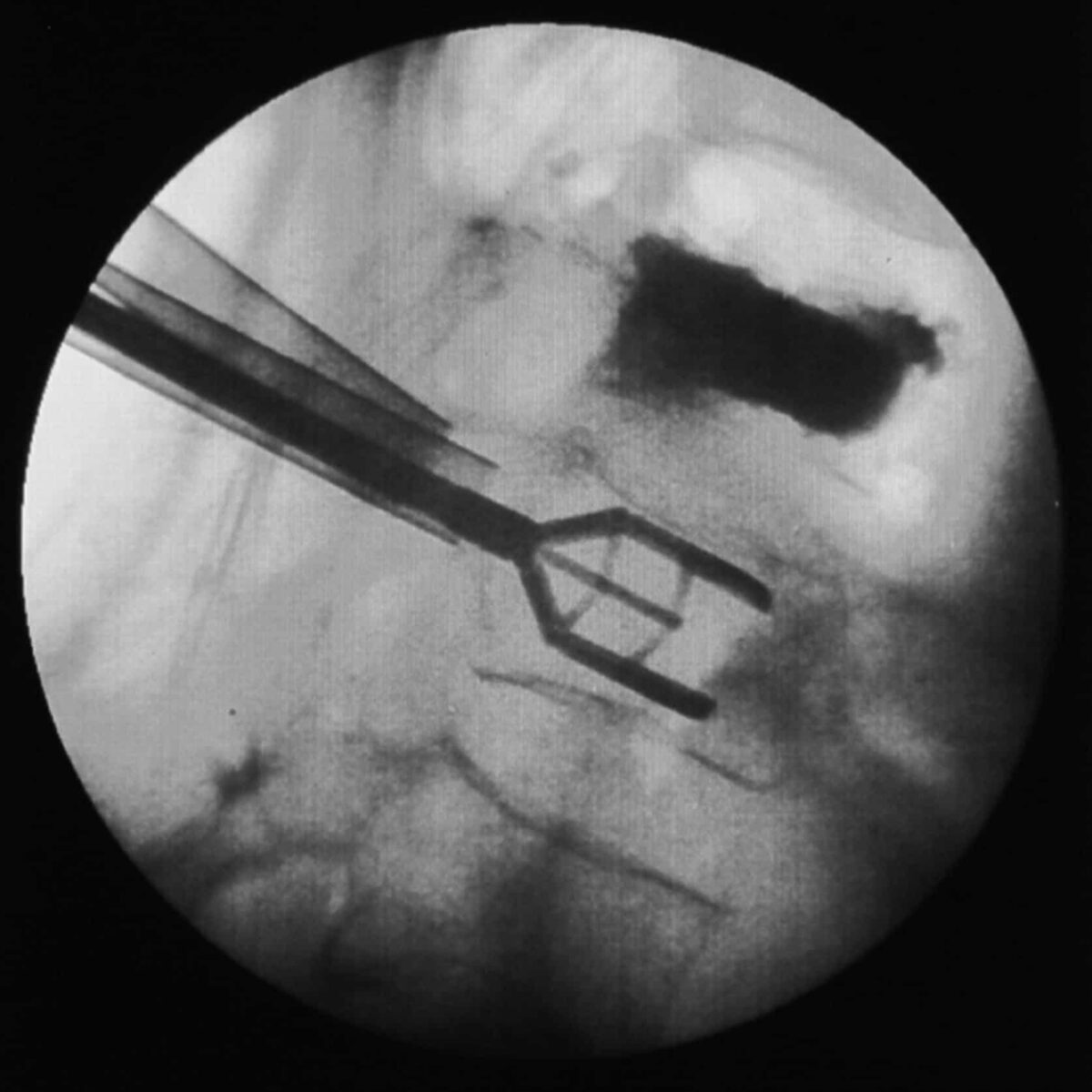
endoscopic-rhizotomy.jpg

Endoscopic Rhizotomy

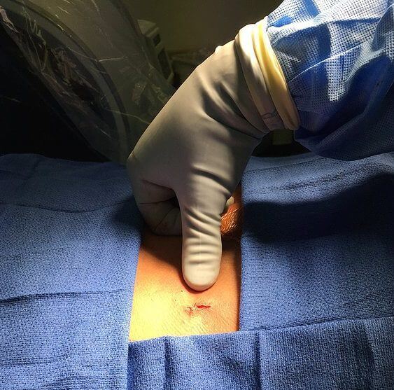
Endoscopic rhizotomy is a minimally invasive endoscopic surgery that allows direct visualization of the medial branch nerve that supplies the facet joints in the back of the spine. The surgery takes the percutaneous RF facet denervation procedure an important step further by providing direct endoscopic visualization of the posterior spinal anatomy and nerves. An incision that is less than a quarter of an inch is made, and a camera is inserted in the spine. By cutting a section of the medial branch nerve, the pain signal is interrupted. This surgery can be performed on the cervical, thoracic, and lumbar spine. It can also be performed on the sacroiliac joint for sacroiliac joint disease.

Medial branch nerves are very small nerves that innervate the facet joints of the spine. Facet joints are the joints connecting the different vertebra of the spine to each other. The joints are present on both sides of the spine from the neck to the lower back.

When is Endoscopic Rhizotomy indicated?

Radiofrequency Rhizotomy is indicated if a diagnostic procedure, called a medial branch block, is successful in confirming the patient’s back pain is originating from the facet joints. For the sacroiliac joint, a successful sacroiliac joint injection is needed.

Medial branch block is a procedure where local anesthetic is directly placed near the medial branch nerve to block the pain signal carried from the facet joints to the brain. It is a diagnostic tool and typically provides only temporary relief from pain. It is critical in assisting spine specialists in diagnosing the specific cause of your back pain.

After the medial branch block your pain may:

  • Go away for a few hours
  • Go away for a few days
  • Not reduce at all

If the pain is relieved after the medial branch block, this indicates that the origin of the pain are the medial branch nerves that were numbed. At that point, we would likely recommend a radiofrequency ablation or an endoscopic rhizotomy to relieve the pain for a longer period of time. With the radiofrequency technique, the nerves regenerate over time and the pain returns after a few months. With the endoscopic technique, a section is cut from the nerve, preventing the nerve from being able to regenerate.


How is an Endoscopic Rhizotomy performed?

Endoscopic rhizotomy is an outpatient, same day, true minimally invasive surgery. During the day of your procedure, you will be taken to the pre-op area where trained nursing staff will get you ready by taking vitals and reviewing your medications. Your blood sugar and coagulation status may also be checked if needed. Then you will enter the operating room where you will lie face down on a table for treatment of the painful area.

The surgery is performed under deep sedation so there is no pain during the surgery. A small (7mm) incision is made in the surgical area and an endoscopic cannula with a camera is inserted into the spine. The doctor is guided by fluoroscopic X-ray to place the camera in the correct position.

The camera allows the surgeon to see the inside of your spine where the nerve usually resides. The surgeon utilizes a microscopic cauterizing instrument to find the small nerve branches that supply the joints in the spine. After identifying the nerve, a section is cut from the nerve, preventing any regrowth in the future.

The camera is removed and the incision is closed with a single absorbable suture that is buried under the skin, so that no suture removal is needed. The procedure takes about 45 minutes to complete but may take longer depending on the number of nerves required to be treated.


What are some possible risk or complications from this minimally invasive surgery?

With the use of careful imaging which allows direct visualization of the spine and spine specialsits trained in the latest endoscopic techniques, complications are very rare. But with all medical procedures, complications may occur. To help minimize risks please follow all directions given to you by your physician. Ensure that all your treatment options are explained so you are aware of the risks and benefits of this surgery.

Some complications may include:

  • Infection: Your surgeon cleans and sterilizes your back before every surgery to prevent this from occurring. This risk is minimized as you are given IV antibiotics one hour prior to the start of surgery.
  • Increased pain may occur after the procedure in the area where the camera was placed. This may last up to a week or rarely longer. However, it typically resolves completely.
  • You may also experience an area of numbness, usually in the area which was originally painful.
  • The procedure may take up to six weeks for complete benefit, so improvement may not be immediate.
    You will need someone to drive you home once you are discharged. As always, follow the instructions of your care provider and have your questions answered prior to the procedure.

endoscopic-discectomy.jpg

What is an Endoscopic Discectomy?

Endoscopic discectomy is a minimally invasive spine surgery performed to treat disc problems that cause back and leg pain. A unique feature of this technique is the technology used to access the spine; through a keyhole (8mm) incision, surgery is performed using a specialized WOLF endoscope.

A WOLF endoscope is a specialized, german engineered, device with a tiny camera at the end. The endoscope delivers the surgeon an excellent view of the disc and related structures through this tiny incision. This enhanced view allows the surgeon to select the part of the disc to be removed that is causing your pain.

Instead of cutting through tissues (eg, muscles, ligaments) to access the spine, a dilator will gently separate soft tissue and are graduated in size to enlarge the operative field.

What are some of the benefits of Endoscopic Discectomy?

Some of the benefits of a true, minimally invasive, WOLF endoscopic discectomy include:

  • Tiny incision
  • Less tissue disruption
  • Less blood loss
  • Less post-operative pain
  • Accelerated recovery
  • Same Day Surgery
  • Can be performed under light sedation (minimizing anesthesia risk)


What conditions can Endoscopic Discectomy help with?

endoscopic discectomy

(DDD), disc herniation, and disc bulge are examples of disc-related problems that may compress or pinch spinal nerves and cause low back, buttock and leg pain. Other neurologic symptoms may include numbness, tingling, and weakness in one or both legs. The goal of endoscopic discectomy is to decompress the nerves, relieve symptoms, and enable the patient to quickly return to regular activities of daily living.

Not every patient with DDD or disc problem requires spine surgery. Dr. Kountis may recommend endoscopic discectomy for the following reasons:

  • Non-operative treatment fails to relieve symptoms
  • Progressive worsening of neurologic symptoms or pain, and/or neurological problems
  • Numbness, loss of function, and weakness (signs of neurologic dysfunction)


How is Endoscopic Discectomy performed?

Endoscopic discectomy is performed under local and/or intravenous anesthesia; the patient is awake during the surgery. The back area is cleansed and prepared for surgery. A small needle is inserted into the disc guided by fluoroscopy; a type of x-ray used during surgery. A tiny skin incision is made and the dilation tube is slipped into place followed by the endoscope.

Special miniature-sized instruments (eg, forceps, rongeurs) are advanced through the endoscope. A YAG laser may be utilized to remove only the damaged disc part and to bind the disc wall (annulus fibrosus). The laser also reduces (shrinks) the size of the disc to decompress nerve roots. The procedure takes about 30-minutes to one-hour per disc. The incision is closed with one suture and a small bandage.


Disclaimer


The information on this web site is not designed to replace a physician’s independent judgement about the appropriateness or risks of a procedure for a given patient. Always consult your doctor about your medical conditions or back problem. Use of the anagenesisspine.com web site is conditional upon your acceptance.


Subscribe


Sign up for newsletter to receive all the news from Anagenesis Spine & Joint Health.


    ® 2017 All Rights Reserved, Dr. Vasilios Kountis.

    Skip to content