Monday - Friday (BY APPT ONLY)Saturday and Sunday - CLOSED718-962-7246admin@anagenesisspine.com78 Todt Hill Rd, Suite 112Staten Island, NY 10314 New York, NY

Latest news

Classic list

Globally incubate standards compliant channels before scalable benefits. Quickly disseminate superior deliverables whereas web-enabled applications.
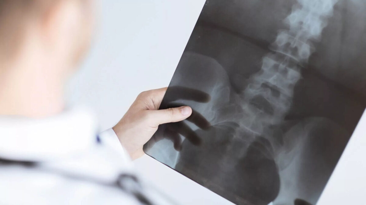
sacroiliac-joint-injection-1200x675.jpg

What is Sacroiliac Joint Pain?

Sacroiliac joint pain or SI joint dysfunction is the result of too much or too little motion occurring at the sacroiliac joint, which leads to inflammation and pain, which can often be debilitating. Part of the pelvis, the SI joint is attached to the front and back by the muscles and ligaments which surround it. Any of these can be the source of pain in a dysfunctional SI joint.


What are some symptoms of Sacroiliac Joint Pain?

Pain can range from dull aching to sharp and stabbing and increases with physical activity. Symptoms also worsen with prolonged or sustained positions (i.e., sitting, standing, lying). Bending forward, stair climbing, hill climbing, and rising from a seated position can also provoke pain. Pain is reported to increase during menstruation in women and sexual intercourse. Some examples include:

  • Lower back & buttock pain
  • Sciatic leg pain
  • Groin pain & hip pain
  • Knee Pain
  • Transient numbness
  • Urinary frequency (rare)


How is Sacroiliac Joint Pain Diagnosed?

Certain physical exam findings correlate with SI joint pain and dysfunction. The current “gold standard” for diagnosis of sacroiliac joint dysfunction emanating within the joint is a sacroiliac joint injection confirmed under fluoroscopy or CT-guidance using a local anesthetic solution.


How do you treat Sacroiliac Joint Pain?

Treatment is often dependent on the duration and severity of the pain and dysfunction. In the acute phase (first one to two weeks) for a mild sprain of the sacroiliac, it is typical for the patient to be prescribed rest, ice/heat, physical therapy, and anti-inflammatory medicine. If the pain does not resolve in the first one to two weeks, then the patient may benefit from a steroid and anesthetic mixture injected via into the joint with fluoroscopic guidance (this also serves in confirming diagnosis).

For the most severe and chronic forms of sacroiliac dysfunction, treatment should proceed with the support of a sacroiliac belt, and a series of prolotherapy injections to aid in regeneration and healing of the surrounding injured ligaments. If that treatment is limited, surgical transection of the sacral lateral branch nerves may be appropriate if pain relief is confirmed with a series of lateral branch blocks confirmed under fluoroscopy.


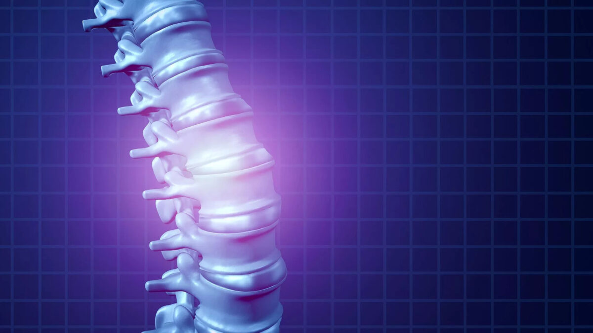
discogram-and-radiofrequency-ablation-1-1200x675.jpg

What is a Radiofrequency Ablation?

Radiofrequency (RF) ablation is a percutaneous procedure offered to select patients with debilitating chronic back pain and joint pain. RF ablation is reserved for select patients with chronic neck or back pain from the spinal joints in the neck or lower back. RF is employed for those patients who fail more conservative therapies. Patients are determined to be appropriate candidates for RF denervation only after failing such back pain treatments and then demonstrating a positive response to a confirmatory diagnostic injection screen.


How do Radiofrequency Ablations work?

The RF technique utilizes heat to denature nerve endings (medial branch nerves) that supply sensation to the affected spinal or peripheral joint. As a result, the joints are inhibited from transmitting pain. This procedure is similar to an injection procedure in that a needle type instrument is utilized and guided with fluoroscopic imaging to reach the target nerve and pain generator. Additional testing during the procedure is performed to ensure proper positioning in the body. As with all of our injection procedures, RF ablation is performed on an outpatient setting with the use of light anesthesia.

Although RF procedures have been increasingly utilized by the spine community over the past decade, the medical literature supporting such use has only recently been established.

Our use of this procedure and procedural technique will be evidence based, and the technology utilized will be the most current to assure patient comfort, minimize procedure time, and maximize clinical outcomes.


What are the Benefits of Radiofrequency Ablation?

The degree of pain relief varies, depending on the cause and location of the pain. Pain relief from RFA can last anywhere from six to 18 months and in some cases, relief can last for years. Typical outcomes will mimic the relief that you experience during your diagnostic medial branch blocks or genicular nerve blocks. In our experience, more than 90 percent of our patients treated with RFA experience significant pain relief.


medial-branch-block-1200x675.jpg

What is a medial branch block?

We offer cervical, thoracic, and lumbar medial branch blocks. This is an outpatient procedure for diagnosing and treating headaches, neck, shoulder, upper back, mid back, lower back, buttock, hip, and groin pain. This information has been provided by your doctor so you can better understand this procedure. Your doctor will make the best recommendation for your specific needs.

During this procedure, a local anesthetic (numbing medicine) is injected near the medial branch nerve. This stops the transmission of pain signals from the facet joint. If your pain is reduced and you are able to move normally, then the doctor will know which facet joints have been causing your pain.


What happens during a medial branch block?

An IV will be inserted to administer intravenous medication(s) to help you relax. A local anesthetic will be used to numb your skin.

Fluoroscopy, a type of x-ray, will be used to insure the safe and proper position of the needle. Once your physician is sure the needle is correctly placed, the medicine will be injected.


What happens after a medial branch block?

You will be monitored for up to 30 minutes after the injection. Before you leave, you will be given discharge instructions. Keeping track of your pain helps the doctor know what the next steps will be. You may want to check for pain by moving in ways that hurt before the injection, but do not overdo it. You may feel immediate pain relief and numbness for a brief period of time after the injection. This means the medication has reached the right spot.

You should be able to return to work the day after the injection, but always check with your doctor.


How long can I expect pain relief?

How long you can expect pain relief depends on how many areas are injured and the amount of inflammation. If your pain goes away for a short time, but then returns, you may be a candidate for radiofrequency ablation (RFA) to the medial branch nerve or a candidate for an endoscopic rhizotomy. This procedure provides a more permanent disruption of pain signals.


facet-joint-injection-1200x675.jpg

What are Facet Joint Injections?

Facet joint injections are safe and effective non-surgical treatments used to relieve pain and inflammation in the facet joints of the spine. The bones of the spine (vertebrae) are connected to each other at the facet joints, which are located at the top and bottom of each vertebra. These joints allow the spine a range of motions. However, conditions like spinal stenosis, sciatica, herniated disc, and arthritis can cause damage to these joints, leading to chronic pain and inflammation. These may also be accompanied by limitation in spine movements, especially in the neck (cervical) and lower back (lumbar) areas. Aside from these areas, pain may be referred to other parts of the body such as the shoulders, upper back, buttocks, hips, groins, or down to the knee. A Chronic Pain Specialist may recommend facet joint injections to manage these symptoms and allow proper healing.


How do Facet Joint Injections Work?

Facet joint injections aim to reduce the inflammation of the joints in the spine. Cervical facet injections reduce neck pain, as well as pain that spreads to the back of the head, shoulder or upper back. Lumbar facet injections, on the other hand, reduce the low back pain, as well as pain that spreads to the buttocks, hips, groins and knees.

Facet joint injections are administered into the lining of the spinal joints where a soft membrane called synovium can become inflamed, causing pain and muscle spasm. The painful area is targeted and a combination of a local anesthetic and an anti-inflammatory drug (steroid) is injected to bathe the affected joint. The treatment has a localized effect, thus avoiding the widespread effects of oral medications. The result is a significant reduction of inflammation and pain over time, which allows one’s muscles to relax and consequently improves one’s mobility for a period of weeks or months. It also facilitates healing and improves a patient’s ability to resume normal activities as well as exercise.


What is the Procedure Like?

Facet joint injections are usually performed as an outpatient procedure by a qualified Chronic Pain Specialist who is an expert with the treatment. After thorough evaluation, the patient is briefed about the procedure and prepared beforehand so that they are relaxed. IV sedation will be provided in order to help you relax and minimize pain during the procedure.

The patient is asked to lie face down on an operating table. The injection site is first sterilized and then numbed using a local anesthetic. Utilizing live x-ray(fluoroscopy) to guide the placement of the needle, your Staten Island doctor targets the affected facet joint and carefully injects the medication. The exact location is confirmed with contrast.

The patient is monitored before they are discharged. They will be asked to evaluate their improvement in the succeeding week and report for follow-up. Repeat injections may be performed up to three times a year if needed.

Depending on the duration of relief, endoscopic rhizotomy may be recommended in order to provide long lasting relief without the need of further injections.


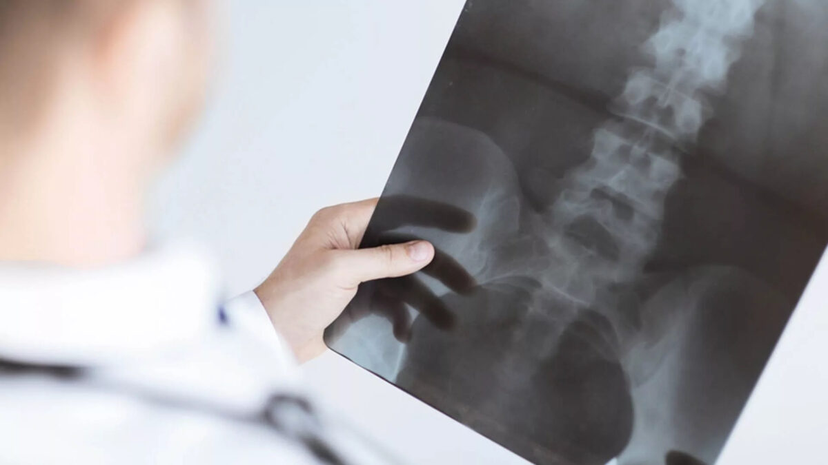
discogram-and-radiofrequency-ablation-1200x675.jpg

What is a Discogram?

A discogram, or diskogram, is a test used to evaluate back pain. A discogram may help your doctor determine if an abnormal disk in your spine is causing your back pain.

Spinal disks look a little like jelly doughnuts, with a tough outer layer and a gel-like substance inside. Disks act as cushions between the bones in your spine.

During a discogram, dye is injected into the soft center of the disk. The injection itself sometimes reproduces your back pain. Several disks may be injected to try to pinpoint the cause of your back pain.

The dye also moves into any cracks in the disk’s exterior, which can then be seen on an X-ray or CT scan. However, disks that show signs of wear and tear don’t always cause symptoms, so the usefulness of a discogram is controversial.


Why is a Discogram done?

A discogram is an invasive test that generally isn’t used for an initial evaluation of back pain. But your doctor may suggest a discogram if your back pain persists despite conservative treatments, such as medication and physical therapy.

Some doctors use a discogram before spinal surgery to help identify which disks need to be treated.


What are the risks?

A discogram is generally a safe procedure. But as with any medical procedure, a discogram carries a rare risk of complications. Possible complications include:

  • Infection
  • Worsening of chronic back pain
  • Headache
  • Injury to nerves in and around the spine
  • Allergic reaction to the dye


How do you prepare for the test?

You may need to avoid taking blood-thinning medications for a period of time before the procedure. Your doctor will give you specific instructions about what medicines you can take. You will need to avoid food or drink the morning before the test.


What should you expect on the day of the procedure?

The total time for the test is about three hours. The discogram itself takes about 30 minutes. You’ll be able to go home later that same day.


What happens during a discogram?

The total time for the test is about three hours. The discogram itself takes about 30 minutes. You’ll be able to go home later that same day.

A discogram is performed in a clinic or center that has imaging equipment. You are awake during the procedure, but your doctor may give you a sedative through a vein to help you relax. You may also receive antibiotics to help prevent infection.

During the procedure, you lie on a table on your abdomen. After cleaning your skin, your doctor may inject a numbing medicine to decrease pain caused by the insertion of the discography needles.

Your doctor will use an imaging technique (fluoroscopy) that enables him or her to watch as the needle enters your body. Fluoroscopy allows more precise and safer placement of the needle into the center of the disk to be examined. A contrast dye is then injected into the disk, and an X-ray or CT scan is taken to see if the dye spreads.

If the dye stays in the center of the disk, the disk is normal. If the dye spreads outside the center of the disk, the disk has undergone some wear-and-tear change. These changes may or may not be the cause of your pain.

Typically, if a disk is causing your back pain, you will feel pain during the injection that’s similar to the back pain you have daily. If a disk is normal, there’s little pain during injection. During discography, you will be asked to rate your pain.


What happens after the discogram?

You remain in the procedure room for approximately 30 minutes to one hour for observation. Someone will need to drive you home.

It is normal to have some pain at the injection site or in the low back for several hours after the procedure. You will need to keep your back dry for 24 hours after the procedure. If you develop severe back pain or you develop a fever one to two weeks after the procedure, call your doctor right away.


Results of a Discogram

Your doctor will review the images and the information you provided about the pain you experienced during the procedure. Both are important to help pinpoint the source of your back pain. Your doctor will use this information to guide your ongoing back pain treatment or prepare for surgery.

Doctors usually don’t rely on the results of a discogram alone to guide treatment. That’s because a disk with wear-and-tear change might not cause pain. Also, pain responses during a discogram can vary widely.

Typically, results of a discogram are combined with results of other tests — such as MRI or CT scan and physical examination — when determining a back pain treatment plan.


lumbar-epidural-steroid-injection-1200x675.jpg

A lumbar epidural steroid injection is an outpatient procedure meant for back pain treatment and leg pain. This information has been provided by your doctor so you can better understand this procedure. Your doctor will make the best recommendation for your specific needs.


What is a lumbar epidural steroid injection?

During this procedure, a corticosteroid (anti-inflammatory medicine) is injected into the epidural space to reduce inflammation and pain. Your physician may inject into the epidural space from behind, this is called an interlaminar injection. When your physician goes in from the side, it is called a transforaminal injection. If the needle is positioned next to an individual nerve root, it is called a selective nerve root block. When performed from below it is called a caudal injection.


What happens during an injection?

An IV is typically inserted to administer intravenous medication(s) to help you relax. A local anesthetic will be used to numb your skin at the area where the procedure will be performed.

Fluoroscopy, a type of x-ray, will be used to ensure the safe and proper position of the needle. A dye will be injected to make sure the needle is in the correct spot and that it flows properly into the epidural space.

The flow around your nerve impingement and herniated disc will be evaluated.

Once your physician is sure the needle is correctly placed, the medicine will be injected.


What happens after an injection?

You will be monitored for up to 30 minutes after the injection. Before you leave, you will be provided with discharge instructions. Keeping track of your pain helps the doctor know what the next steps will be.

It is possible that your pain may become a little worse for a day or two. This is normal. It may be caused by needle irritation, by the steroid itself or the anesthetic wearing off. Steroids usually take two or three days to start working, but can take as long as a week.

You should be able to return to work the day after the injection, but always consult with your doctor.


How long can I expect pain relief?

The amount and duration of pain relief varies from person to person and is dependent on many factors including your underlying pathology and activity level.

Some can have relief that lasts for years, while others may have short-term relief. Usually a series of injections, often three, each spaced two weeks apart, are given. It is important to discuss with your physician your response to epidural steroids in order to plan future treatment options.


cervical-epidural-injection-1200x675.jpg

What is a cervical epidural steroid injection?

A cervical epidural steroid injection is an outpatient procedure for treating neck, upper back, shoulder, and arm pain. This information has been provided by your doctor so you can better understand this procedure. Your doctor will make the best recommendation for your specific needs.

During this procedure, a corticosteroid (anti-inflammatory medicine) is injected into the epidural space to reduce inflammation and pain. Your physician may inject into the epidural space from behind; this is called an interlaminar injection. When your physician goes in from the side, it is called a transforaminal injection. If the needle is positioned next to an individual nerve root, it is called a selective nerve root block.


What happens during an injection?

An IV will be inserted to administer intravenous medication(s) to help you relax. A local anesthetic will be used to numb your skin.

Fluoroscopy, a type of x-ray, will be used to ensure the safe and proper position of the needle. A contrast dye will also be injected to make sure the needle is in the correct spot.

Once your physician is sure the needle is correctly placed, the medicine will be injected.

Your pain may become a little worse for a day or two. This is normal. It may be caused by needle irritation or by the steroid itself. Steroids usually take two or three days to start working, but can take as long as a week.

You should be able to return to work the day after the injection, but always check with your doctor.


How long can I expect pain relief?

The amount and duration of pain relief varies from person to person and is dependent on many factors including underlying pathology and activity level. Some can have relief that lasts for years, while others have short-term relief.

Usually a series of cervical epidural steroid injections, often three, each spaced a week or two apart, are given. It is important to discuss with your physician your response to epidural steroids in order to plan future treatment options.


Disclaimer


The information on this web site is not designed to replace a physician’s independent judgement about the appropriateness or risks of a procedure for a given patient. Always consult your doctor about your medical conditions or back problem. Use of the anagenesisspine.com web site is conditional upon your acceptance.


Subscribe


Sign up for newsletter to receive all the news from Anagenesis Spine & Joint Health.


    ® 2017 All Rights Reserved, Dr. Vasilios Kountis.

    Skip to content