Monday - Friday (BY APPT ONLY)Saturday and Sunday - CLOSED718-962-7246admin@anagenesisspine.com78 Todt Hill Rd, Suite 112Staten Island, NY 10314 New York, NY

Latest news

Classic list

Globally incubate standards compliant channels before scalable benefits. Quickly disseminate superior deliverables whereas web-enabled applications.
discogram-and-radiofrequency-ablation-1200x675.jpg

What is a Discogram?

A discogram, or diskogram, is a test used to evaluate back pain. A discogram may help your doctor determine if an abnormal disk in your spine is causing your back pain.

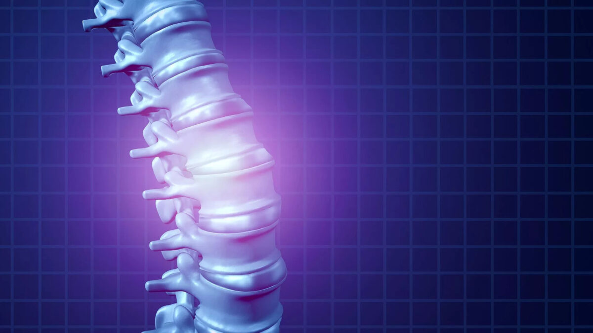
Spinal disks look a little like jelly doughnuts, with a tough outer layer and a gel-like substance inside. Disks act as cushions between the bones in your spine.

During a discogram, dye is injected into the soft center of the disk. The injection itself sometimes reproduces your back pain. Several disks may be injected to try to pinpoint the cause of your back pain.

The dye also moves into any cracks in the disk’s exterior, which can then be seen on an X-ray or CT scan. However, disks that show signs of wear and tear don’t always cause symptoms, so the usefulness of a discogram is controversial.


Why is a Discogram done?

A discogram is an invasive test that generally isn’t used for an initial evaluation of back pain. But your doctor may suggest a discogram if your back pain persists despite conservative treatments, such as medication and physical therapy.

Some doctors use a discogram before spinal surgery to help identify which disks need to be treated.


What are the risks?

A discogram is generally a safe procedure. But as with any medical procedure, a discogram carries a rare risk of complications. Possible complications include:

  • Infection
  • Worsening of chronic back pain
  • Headache
  • Injury to nerves in and around the spine
  • Allergic reaction to the dye


How do you prepare for the test?

You may need to avoid taking blood-thinning medications for a period of time before the procedure. Your doctor will give you specific instructions about what medicines you can take. You will need to avoid food or drink the morning before the test.


What should you expect on the day of the procedure?

The total time for the test is about three hours. The discogram itself takes about 30 minutes. You’ll be able to go home later that same day.


What happens during a discogram?

The total time for the test is about three hours. The discogram itself takes about 30 minutes. You’ll be able to go home later that same day.

A discogram is performed in a clinic or center that has imaging equipment. You are awake during the procedure, but your doctor may give you a sedative through a vein to help you relax. You may also receive antibiotics to help prevent infection.

During the procedure, you lie on a table on your abdomen. After cleaning your skin, your doctor may inject a numbing medicine to decrease pain caused by the insertion of the discography needles.

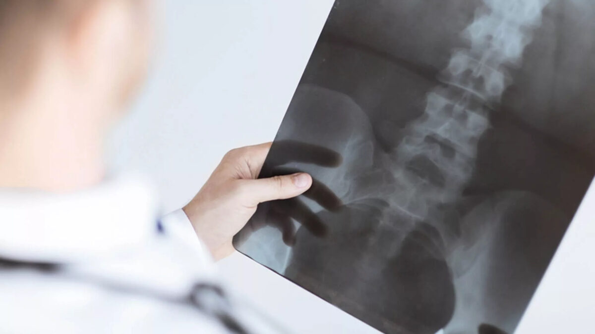
Your doctor will use an imaging technique (fluoroscopy) that enables him or her to watch as the needle enters your body. Fluoroscopy allows more precise and safer placement of the needle into the center of the disk to be examined. A contrast dye is then injected into the disk, and an X-ray or CT scan is taken to see if the dye spreads.

If the dye stays in the center of the disk, the disk is normal. If the dye spreads outside the center of the disk, the disk has undergone some wear-and-tear change. These changes may or may not be the cause of your pain.

Typically, if a disk is causing your back pain, you will feel pain during the injection that’s similar to the back pain you have daily. If a disk is normal, there’s little pain during injection. During discography, you will be asked to rate your pain.


What happens after the discogram?

You remain in the procedure room for approximately 30 minutes to one hour for observation. Someone will need to drive you home.

It is normal to have some pain at the injection site or in the low back for several hours after the procedure. You will need to keep your back dry for 24 hours after the procedure. If you develop severe back pain or you develop a fever one to two weeks after the procedure, call your doctor right away.


Results of a Discogram

Your doctor will review the images and the information you provided about the pain you experienced during the procedure. Both are important to help pinpoint the source of your back pain. Your doctor will use this information to guide your ongoing back pain treatment or prepare for surgery.

Doctors usually don’t rely on the results of a discogram alone to guide treatment. That’s because a disk with wear-and-tear change might not cause pain. Also, pain responses during a discogram can vary widely.

Typically, results of a discogram are combined with results of other tests — such as MRI or CT scan and physical examination — when determining a back pain treatment plan.


lumbar-epidural-steroid-injection-1200x675.jpg

A lumbar epidural steroid injection is an outpatient procedure meant for back pain treatment and leg pain. This information has been provided by your doctor so you can better understand this procedure. Your doctor will make the best recommendation for your specific needs.


What is a lumbar epidural steroid injection?

During this procedure, a corticosteroid (anti-inflammatory medicine) is injected into the epidural space to reduce inflammation and pain. Your physician may inject into the epidural space from behind, this is called an interlaminar injection. When your physician goes in from the side, it is called a transforaminal injection. If the needle is positioned next to an individual nerve root, it is called a selective nerve root block. When performed from below it is called a caudal injection.


What happens during an injection?

An IV is typically inserted to administer intravenous medication(s) to help you relax. A local anesthetic will be used to numb your skin at the area where the procedure will be performed.

Fluoroscopy, a type of x-ray, will be used to ensure the safe and proper position of the needle. A dye will be injected to make sure the needle is in the correct spot and that it flows properly into the epidural space.

The flow around your nerve impingement and herniated disc will be evaluated.

Once your physician is sure the needle is correctly placed, the medicine will be injected.


What happens after an injection?

You will be monitored for up to 30 minutes after the injection. Before you leave, you will be provided with discharge instructions. Keeping track of your pain helps the doctor know what the next steps will be.

It is possible that your pain may become a little worse for a day or two. This is normal. It may be caused by needle irritation, by the steroid itself or the anesthetic wearing off. Steroids usually take two or three days to start working, but can take as long as a week.

You should be able to return to work the day after the injection, but always consult with your doctor.


How long can I expect pain relief?

The amount and duration of pain relief varies from person to person and is dependent on many factors including your underlying pathology and activity level.

Some can have relief that lasts for years, while others may have short-term relief. Usually a series of injections, often three, each spaced two weeks apart, are given. It is important to discuss with your physician your response to epidural steroids in order to plan future treatment options.


cervical-epidural-injection-1200x675.jpg

What is a cervical epidural steroid injection?

A cervical epidural steroid injection is an outpatient procedure for treating neck, upper back, shoulder, and arm pain. This information has been provided by your doctor so you can better understand this procedure. Your doctor will make the best recommendation for your specific needs.

During this procedure, a corticosteroid (anti-inflammatory medicine) is injected into the epidural space to reduce inflammation and pain. Your physician may inject into the epidural space from behind; this is called an interlaminar injection. When your physician goes in from the side, it is called a transforaminal injection. If the needle is positioned next to an individual nerve root, it is called a selective nerve root block.


What happens during an injection?

An IV will be inserted to administer intravenous medication(s) to help you relax. A local anesthetic will be used to numb your skin.

Fluoroscopy, a type of x-ray, will be used to ensure the safe and proper position of the needle. A contrast dye will also be injected to make sure the needle is in the correct spot.

Once your physician is sure the needle is correctly placed, the medicine will be injected.

Your pain may become a little worse for a day or two. This is normal. It may be caused by needle irritation or by the steroid itself. Steroids usually take two or three days to start working, but can take as long as a week.

You should be able to return to work the day after the injection, but always check with your doctor.


How long can I expect pain relief?

The amount and duration of pain relief varies from person to person and is dependent on many factors including underlying pathology and activity level. Some can have relief that lasts for years, while others have short-term relief.

Usually a series of cervical epidural steroid injections, often three, each spaced a week or two apart, are given. It is important to discuss with your physician your response to epidural steroids in order to plan future treatment options.


spinal-stenosis-1200x675.jpg

What is Spinal Stenosis?

Spinal Stenosis is a narrowing of the canal of the spine that can cause pressure to be placed on the spinal cord and nerves of the spine. In a healthy spinal column, nerve roots leave the spine via nerve root canals unrestricted. Spinal stenosis occurs when bone and soft tissues begin to grow over top of the vertebrae and end up covering the nerve root canals.

The most common types of spinal stenosis are Lumbar Spinal Stenosis and Cervical Spinal Stenosis. Lumbar Spinal Stenosis is more common while Cervical Spinal Stenosis is more dangerous.

The most common cause of Spinal Stenosis is a natural wear and tear due to the aging process in which ligaments may thicken, herniated discs or bulging discs may occur and cysts may form. Arthritis and spinal injuries are also seen as causes for Spinal Stenosis.


What are the symptoms of Spinal Stenosis?

It is possible to have Spinal Stenosis and have no signs or symptoms of having the condition. In those who do have symptoms, they usually are gradual symptoms and may become worse over time. Spinal Stenosis causes constriction to the nerve roots and can lead to a number of painful and unpleasant symptoms as a result. The most common symptoms of Spinal Stenosis include:

  • Lower back pain
  • Leg cramping
  • Leg discomfort
  • Numbness, weakness or tingling in your leg, foot, hand or arm
  • Decreased physical activity


Cervical Radiculopathy Treatment

At Anagenesis Spine & Pain Medicine, personalized treatment plans are designed for each individual patient by our expert staff. Common treatments for Spinal Stenosis at Staten Island Pain Care include:

  • Physical Therapy
  • Epidural Steroid Injections
  • Platelet Rich Plasma Injections
  • Stem Cell Injections
  • Surgical Decompression

facet-joint-syndrome-1200x675.jpg

What is Facet Joint Syndrome?

Facet Joint Syndrome is when the facet joints either weaken or become stressed and damaged. Several aspects of our everyday life cause this ailment; from everyday wear and tear to a specific injury to the back or neck and degeneration of a disc. Cartilage eventually wears away from protecting the facet joints, which can cause a variety of symptoms depending upon which region of the spine is damaged.


Why does my back hurt all of a sudden?

Facet joints are highly sensitive to irritation. They’re designed to slide around on each other. There is a tiny fluid-filled capsule on each facet that keeps the joints lubricated. These sacs house tiny nerve endings that let you know when they’re not happy. You’ll feel facet joint paint if they’re injured because the pain is persistent. You can either stop what you’re doing or seek help from a pain management doctor.

Facet joints are working all the time, so they do wear out. When that happens, bone spurs form, protruding into your nerves or the joints themselves become arthritic. When joints degenerate, you experience severe pain whenever you’re in motion. That’s when you need to visit Dr. Kountis, a Staten Island Spine Specialist for treatment options.


What are some treatment options for Facet Joint Syndrome in Staten Island?

Long-term facet joint pain treatment involves exercise, strict attention to your posture, alternating hot and cold applications and changing certain activities that lead to pain. Anti-inflammatories, manual manipulation, and the right kind of pillows help beat back the pain. A procedure that freezes the ends of the nerves is an option, and your doctor should discuss the implications of this treatment with you.

Bone fusion surgery is the last resort your pain management doctor may consider — and only after your pain consistently remains severe and debilitating.


sacroiliac-joint-pain-1200x675.jpg

What is Sacroiliac Joint Pain?

Sacroiliac joint pain or SI joint dysfunction is the result of too much or too little motion occurring at the sacroiliac joint, which leads to inflammation and pain, which can often be debilitating. Part of the pelvis, the SI joint is attached to the front and back by the muscles and ligaments which surround it.

Any of these can be the source of pain in a dysfunctional SI joint.


What are some symptoms of Sacroiliac Joint Pain?

While it is not clear how the pain is caused, it is thought that an alteration in the normal joint motion may be the culprit that causes sacroiliac joint pain. This source of pain can be caused by either:

  • Too much movement (hypermobility or instability): The pain is typically felt in the lower back and/or hip and may radiate into groin area.
  • Too little movement (hypomobility or fixation):
    • The pain is typically felt on one side of the low back or buttocks, and can radiate down the leg.
    • The pain usually remains above the knee, but at times pain can extend to the ankle or foot.
    • The pain is similar to sciatica, or pain that radiates down the sciatic nerve and is caused by a radiculopathy.
  • This condition is generally more common in young and middle-aged women.


How is Sacroiliac Joint Pain Diagnosed?

Certain physical exam findings correlate with SI joint pain and dysfunction. The current “gold standard” for diagnosis of sacroiliac joint dysfunction emanating within the joint is sacroiliac joint injection confirmed under fluoroscopy or CT-guidance using a local anesthetic solution.


Treatment for Sacroiliac Joint Pain

Treatment is often dependent on the duration and severity of the pain and dysfunction. In the acute phase (first 1–2 weeks) for a mild sprain of the sacroiliac, it is typical for the patient to be prescribed rest, ice/heat, physical therapy, and anti-inflammatory medicine.

If the pain does not resolve in the first 1–2 weeks, then the patient may benefit from a steroid and anesthetic mixture injected via ultrasound guidance into the joint (this also serves in confirming diagnosis), as well as manipulative or manual therapy.

For the most severe and chronic forms of sacroiliac dysfunction, treatment should proceed with the support of a sacroiliac belt, and a series of prolotherapy injections to aid in regeneration and healing of the surrounding injured ligaments.


Getting Sciatica Treatment in Staten Island

In addition to conservative care, our Sciatica doctors in Staten Island specialize in treating your sciatica in a precise yet effective manner. These cutting edge therapies and treatment options will allow you to return to work and improve your life. Additional manual therapies, electrical stimulation, ultrasound therapy and pain management options are available. Request a consultation to meet with one of our Top Sciatica Specialists.

Call us today at 212-235-1265 or schedule online for the next available appointment to be evaluated by one of Staten Island top sciatica doctors!


cervical-radiculopathy-1200x675.jpg

What is Cervical Radiculopathy?

Cervical radiculopathy, commonly called a “pinched nerve” occurs when a nerve in the neck is compressed or irritated where it branches away from the spinal cord. This may cause pain that radiates into the shoulder, as well as muscle weakness and numbness that travels down the arm and into the hand.

Cervical radiculopathy is often caused by “wear and tear” changes that occur in the spine as we age, such as arthritis. In younger people, it is most often caused by a sudden injury that results in a herniated disk.

In most cases, cervical radiculopathy responds well to conservative treatment that includes medication and physical therapy.


Causes of Cervical Radiculopathy

Any condition that somehow compresses or irritates a cervical nerve can cause cervical radiculopathy. The most common causes include:

Cervical Herniated Disc. If the inner material of the cervical disc leaks out and inflames or impinges the adjacent nerve, it can cause cervical radiculopathy. If a younger person (20s or 30s) has cervical radiculopathy, the most likely cause is a herniated disc.

Cervical Spinal Stenosis. As part of the degenerative process of the cervical spine, changes in the spinal joints can lead to tightening of the space for the spinal canal. Spinal stenosis is a common cause of cervical radiculopathy symptoms in people over age 60.

Cervical Degenerative Disc Disease. When a disc in the cervical spine degenerates, the disc becomes flatter and stiffer and does not support the spine as well. In some people this degenerative process can lead to inflammation or impingement of the nearby nerve root. Cervical degenerative disc disease is a common cause of radiculopathy in people over age 50.


Cervical Radiculopathy Treatment

It is important to note that the majority of patients with cervical radiculopathy get better over time and do not need treatment. For some patients, the pain goes away relatively quickly—in days or weeks. For others, it may take longer.

It is also common for cervical radiculopathy that has improved to return at some point in the future. Even when this occurs, it usually gets better without any specific treatment.

In some cases, cervical radiculopathy does not improve, however. These patients require evaluation and treatment.


Nonsurgical Cervical Radiculopathy Treatment

Initial treatment for cervical radiculopathy is nonsurgical. Nonsurgical treatment options include:

Soft cervical collar. This is a padded ring that wraps around the neck and is held in place with Velcro. Your doctor may advise you to wear a soft cervical collar to allow the muscles in your neck to rest and to limit neck motion. This can help decrease the pinching of the nerve roots that accompany movement of the neck. A soft collar should only be worn for a short period of time since long-term wear may decrease the strength of the muscles in your neck.

Physical therapy. Specific exercises can help relieve pain, strengthen neck muscles, and improve range of motion. In some cases, traction can be used to gently stretch the joints and muscles of the neck.

Medications. In some cases, medications can help improve your symptoms.

Nonsteroidal anti-inflammatory drugs (NSAIDs). NSAIDs, including aspirin, ibuprofen, and naproxen, may provide relief if your pain is caused by nerve irritation or inflammation.

Oral corticosteroids. A short course of oral corticosteroids may help relieve pain by reducing swelling and inflammation around the nerve.

Epidural Steroid injection. In this procedure, steroids are injected near the affected nerve to reduce local inflammation. The injection may be placed between the laminae (epidural injection) or in the foramen (selective nerve injection). Although steroid injections do not relieve the pressure on the nerve caused by a narrow foramen or by a bulging or herniated disk, they may lessen the swelling and relieve the pain long enough to allow the nerve to recover.

Narcotics. These medications are reserved for patients with severe pain that is not relieved by other options. Narcotics are usually prescribed for a limited time only.


Surgical Treatment

If after a period of time nonsurgical treatment does not relieve your symptoms, your doctor may recommend surgery. There are several surgical procedures to treat cervical radiculopathy. The procedure your doctor recommends will depend on many factors, including what symptoms you are experiencing and the location of the involved nerve root.


sciatica-1200x675.jpg

What is Sciatica?

Sciatica, or Lumbar Radiculopathy, refers to lower back pain that radiates down to the leg due to compression of the sciatic nerve. While Sciatica is very common, it is important to work with a sciatica doctor in Staten Island to understand the root cause of it to prevent worsening sciatica pain and flare ups in the future.


What is The Cause of Sciatica?

Sciatica is a symptom associated with multiple spine conditions. Often, Sciatica is a result of a spinal disc which has protruded from its normal position in the vertebral column and is placing pressure on the radicular nerve (nerve root) in the lower back. Herniated discs, spinal stenosis and degenerative disc disease are underlying spine conditions issues that are also associated with Sciatica pain.


What are The Symptoms of Sciatica?

For some individuals sciatica pain may be bearable yet irritating, while for others it may be severe and debilitating.

Symptoms associated with Sciatica are typically one or more of the following:

  • Low back pain
  • Pain on one side of the buttock or in one leg that becomes worse when sitting
  • Burning or tingling down the leg
  • Weakness, numbness, or difficulty in moving the leg or foot
  • A constant pain on one side of the posterior
  • A sharp pain that may make it difficult to stand or walk


Sciatica Treatment in Staten Island

Fortunately, sciatica pain can often be alleviated using our non-surgical treatment methods at our Staten Island Clinic. Patients typically see relief and improvement of sciatica pain symptoms within a few weeks to months after beginning non-surgical sciatica treatment in Staten Island. In patients with severe nerve pain, surgery may be necessary, but our goal is to help you avoid it.

Consulting immediately with a sciatica doctor in Staten Island is recommended for individuals experiencing pain symptoms that might be associated with Sciatica. By diagnosing the area and degree of pain, our sciatica doctors can provide the right level of treatment to help you heal quicker.


Getting Sciatica Treatment in Staten Island

In addition to conservative care, our Sciatica doctors in Staten Island specialize in treating your sciatica in a precise yet effective manner. These cutting edge therapies and treatment options will allow you to return to work and improve your life. Additional manual therapies, electrical stimulation, ultrasound therapy and pain management options are available. Request a consultation to meet with one of our Top Sciatica Specialists.

Call us today at 212-235-1265 or schedule online for the next available appointment to be evaluated by one of Staten Island top sciatica doctors!


herniated-discs-1200x675.jpg

What is a Herniated Disc?

Normal discs in your back function as rubbery cushions between the vertebrae (bones), which make up the spine. These discs act as natural shock absorbers between the vertebrae. When a disc becomes damaged due to a tear, a crack or a rupture, it can cause compression of the spinal nerves. This is what we call a disc herniation.

Herniated discs in the neck cause neck pain and are called cervical disc herniations. Herniated discs in the lower back are called lumbar spine disc herniations. These are the two most common parts of the spine to be affected by a herniated disc. Our herniated disc doctor, Dr. Kountis, is an expert in providing herniated disc treatments in Staten Island.


What causes a Herniated Disc?

Herniated discs are common and can be caused by hard falls, direct impact, sports injuries and age. As we age, discs become dry and brittle, leaving them more susceptible to tearing, cracks, and injury.

Infographic about herniated discs


What are the symptoms of a Herniated Disc?

Symptoms of a herniated disc vary depending on the location in the spine of the affected disc.


Common symptoms of a cervical disc herniation include:

  • Neck pain
  • Pain, weakness or numbing of the shoulders and/or arms
  • Tingling of the arm


Common symptoms of a lumbar disc herniation include:

  • Low back pain
  • Pain, weakness of numbing of the buttocks or leg (sciatica)
  • Tingling of the leg
  • Difficulty and pain walking or standing for long periods of time


Herniated Disc Treatment in Staten Island

Prior to treatment for a herniated disc, Dr. Kountis will ask for a history of the issue, provide a physical examination and may request an MRI. Fortunately, if you do in fact have a herniated disc, our herniated disc doctor specializes in non-surgical and minimally invasive treatments to help relieve your pain and improve function.

The Staten Island herniated disc doctor at Anagenesis Spine & Pain Medicine will tailor a treatment plan to meet your individual needs. Treatments in Staten Island may include physical therapy, acupuncture, manual therapies, epidural steroid injections and nerve root blocks.


degenerative-disc-disease-1200x675.jpg

What is Degenerative Disc Disease?

One of the most common causes of back pain in New York and nationwide is known as degenerative disc disease. As individuals age or experience a significant injury to their back, the spinal discs weaken and can cause sections of the vertebrae to rub together and press on nerves causing them to become pinched. While degenerative disc disease isn’t actually a disease, it is the medical description for common conditions pain management specialists often treat.


What are symptoms of Degenerative Disc Disease?

Inflammation and abnormal micromotion instability are two symptoms associated with degenerative disc disease. Proteins in the disc space may cause inflammation, leading to low back pain radiating to the hips. Pain might also travel down the back of the legs as well. If the outer rings of the intervertebral disc are damaged or become worn down, it is not as effective in resisting motion in the spine. This condition is known as micromotion instability. Both inflammation and abnormal micromotion instability cause lower back spasms and other issues.

There are a number of lower back pain treatments for degenerative disc disease. For expert care and treatment in the Staten Island region, consult with Dr. Vasilios Kountis.


What Happens with Degenerative Disc Disease?

Degenerative disc disease first occurs as small tears appear in the disc wall causing pain. As they heal, they form scar tissue, which is not as strong as the previous material in the disc wall. Over time, this tissue can weaken, causing the disc center to eventually collapse. Bone spurs can then form from the awkward positioning of the vertebrae. Nerve roots can often become compressed and cause severe pain that may radiate down the leg as well.


What are some of the leading treatments for Degenerative Disc Disease?

Once this diagnosis is confirmed by history and physical examination there are a multitude of treatment options available. Typically oral analgesics are trialed which may include medicine like Naprosyn, Ibuprofen or Meloxicam. Of these three Meloxicam tends to have the most favorable profile and is the most convenient with dosing that is once daily.

Treatment options include targeted physical therapy for spinal stabilization exercises and improvement in your overall mobility. We must not forget that the goal of any treatment should be to relieve pain and improve quality of life and functional ability!

If oral analgesics and at least a six-week course of physical therapy does not provide significant relief, medial branch blocks of the cervical and/or lumbar spine are indicated. This diagnostic procedure will pinpoint which part of the spine is primarily responsible for most of the axial back pain. Once confirmed, radio-frequency ablation or endoscopic rhizotomy would likely be warranted.

Patients with signs of degenerative disc disease in its earliest stage have additional preventative treatments with regenerative options. There is significant literature in the medical community that demonstrate intradiskal PRP to decrease pain and the progression of arthritis in the spine. If we can avoid unnecessary surgery (fusion of the spine) by halting the progression of a debilitating disease we should always keep that first and foremost!


Disclaimer


The information on this web site is not designed to replace a physician’s independent judgement about the appropriateness or risks of a procedure for a given patient. Always consult your doctor about your medical conditions or back problem. Use of the anagenesisspine.com web site is conditional upon your acceptance.


Subscribe


Sign up for newsletter to receive all the news from Anagenesis Spine & Joint Health.


    ® 2017 All Rights Reserved, Dr. Vasilios Kountis.

    Skip to content